Thyroid Nodule Image Joint Segmentation and Classification Based on Deep Learning
DOI:
https://doi.org/10.21271/ZJPAS.35.5.6Keywords:
Thyroid nodule; VGG-SegNet; Deep Learning; VGG 19; Fuzzy co-occurrence matrix.Abstract
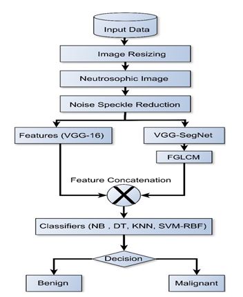
A thyroid nodule is a thyroid gland condition that must be diagnosed and treated as soon as possible to mitigate the threat of death in a possible patient. This research proposed a joint segmentation and classification system to detect and identify thyroid nodules in ultrasound images automatically. The proposed scheme as it is envisioned has consist of two stages: in the first stage thyroid image features were calculated via a concatenation of features vector (to enhance the diversity of image features)generated by deep learning-based VGG-19 model in the first place and the second place by deriving handmade features from VGG-SegNet image segmentation model followed by computing a Fuzzy Grey Level Co-Occurrence Matrix (FGLCM) for each segmented image, which has the effect of eliminating directional difference by multi-angle fusing the grey level co-occurrence matrix (GLCM) and calculation of the membership of each pixel to the texture unit after applying the fuzzy c-means algorithm to the grey level co-occurrence matrix. The second stage then involves thyroid image classification based on four types of machine learning techniques namely (Naïve Bayes NB, Decision Tree DT, K-Nearest Neighbor KNN, and SVM-RBF Support Vector Machine based Radial Basis Function). The proposed model has been evaluated based on Thyroid Digital Image Database (TDID), which is a public dataset for thyroid nodule segmentation created by Universidad Nacional de Colombia. The experimental results revealed that the SVM-RBF classifier has achieved a validation accuracy of 99.25% with concatenated features vector.
References
Acharya, U. R., Chowriappa, P., Fujita, P. et al. 2016 Thyroid lesion classification in 242 patient population using Gabor transform features from high-resolution ultrasound images, Knowledge-Based Systems, 107, pp. 235–245. doi: 10.1016 /j. knosys. 2016.06.010.
Adrian, R. (2017), Image Net: VGGNET, ResNet, Inception, and Xception with Keras. Available at:
https://www.pyimagesearch.com/2017/03/20/imagenet-vggnet-resnet-inception-xception-keras/ . (Accessed: 20 March 2020).
Ajilisa, O.A., Jagathy Raj, V.P., and Sabu, M.K. 2022. Segmentation of Thyroid Nodules from Ultrasound Images Using Convolutional Neural Network Architectures. 687 – 705.
Alom, M.Z., Taha, T.M., Yakopcic, C., Westberg, S., Sidike, P., Nasrin, M.S., Van Esesn, B.C., Awwal, A.A.S. and Asari, V.K., 2018. The history began from alexnet: A comprehensive survey on deep learning approaches. arXiv preprint arXiv:1803.01164.
Cheng HD, Shan J, Ju W, Guo Y, Zhang L. 2010. Automated breast cancer detection and classification using ultrasound images: A survey. Pattern Recognit. 43(1):299-317. doi:10.1016/j.patcog.2009.05.012
Ding, J., Z. Huang, M. Shi and C. Ning. 2019. Automatic Thyroid Ultrasound Image Segmentation Based on U-shaped Network. 12th International Congress on Image and Signal Processing, BioMedical Engineering and Informatics (CISP-BMEI).
Garg A, Tandon N, Varde A. 2020. I am guessing you can’t recognize this: generating adversarial images for object detection using spatial commonsense (student abstract). Proc AAAI Conf Artif Intell 34(10):13789–13790. https://doi.org/10.1609/aaai.v34i10.7166.
Haji, S. O., and R. Z. Yousif. 2019. A Novel Neutrosophic Method for Automatic Seed Point Selection in Thyroid Nodule Images. BioMed Research International. 1, 1-14.
Haji, S.O. and Yousif, R.Z., 2019. A novel run-length based wavelet features for screening thyroid nodule malignancy. Brazilian Archives of Biology and Technology, 62.
Karthikeyan D, Varde AS, Wang W. 2020).Transfer learning for decision support in Covid-19 detection from a few images in big data. IEEE Int Conf Big Data (big Data)4873–4881. https://doi. org/10.1109/BigData50022.2020.9377886.
Kataoka H, Iwata K, Satoh Y. 2015. Feature evaluation of deep convolutional neural networks for object recognition and detection. https://arxiv.org/abs/1509.07627
Kim, K.; Chon, N.; Jeong, H.-W.; Lee, Y. 2022. Improvement of Ultrasound Image Quality Using Non-Local Means Noise-Reduction Approach for Precise Quality Control and Accurate Diagnosis of Thyroid Nodules. Int. J. Environ. Res. Public Health, 19, 13743. https://doi.org/10.3390/ijerph192113743
Koundal, D., Gupta, S. and Singh, S. 2018. Biomedical Signal Processing and Control Computer-aided thyroid nodule detection system using medical ultrasound images’, Biomedical Signal Processing and Control. Elsevier Ltd, 40, pp. 117–130. doi: 10.1016/j.bspc.2017.08.025.
Li, X., Zhang, S., Zhang, Q., Wei, X., Pan, Y., Zhao, J., Xin, X., Qin, C., Wang, X., Li, J.; et al.2019. Diagnosis of thyroid cancer using deep convolutional neural network models applied to sonographic images: A retrospective, multicohort, diagnostic study. Lancet Oncol. 20, 193–201. [CrossRef]
Ma, J. L., F. Wu, T. A. Jiang, Q. Y. Zhao and D. X. Kong. 2017. Ultrasound image-based thyroid nodule automatic segmentation using convolutional neural networks. International Journal of Computer Assisted Radiology and Surgery 12(11):1895-1910, https://doi.org/10.1007/s11548-017-1649-7
Mascarenhas, S. and Agarwal, M., 2021, November. A comparison between VGG16, VGG19 and ResNet50 architecture frameworks for Image Classification. In 2021 International Conference on Disruptive Technologies for Multi-Disciplinary Research and Applications 1(1), pp. 96-99.
Mohammed, B.N., Al-Mukhtar, F.H., Yousif, R.Z. and Almashhadani, Y.S., 2021. Automatic Classification of Covid-19 Chest X-Ray Images Using Local Binary Pattern and Binary Particle Swarm Optimization for Feature Selection. Cihan University-Erbil Scientific Journal, 5(2), pp.46-51.
Nguyen, D.T., Kang, J.K., Pham, T.D., Batchuluun, G., Park, K.R.2022. Ultrasound Image-Based Diagnosis of Malignant Thyroid Nodule Using Artificial Intelligence. Sensors, 20, 1822. [CrossRef]
Pedraza, L., Vargas, C., Narváez, F., Durán, O., Muñoz, E., Romero, E. 2015. An open-access thyroid ultrasound image database. In Proceedings of the 10th International Symposium on Medical Information Processing and Analysis, Cartagena de Indias, Colombia, 28 January; Volume 9287, pp. 1–6.
Prochazka, A., Gulati, S., Holinka, S., Smutek, D. 2019. Classification of Thyroid Nodules in Ultrasound Images Using DirectionIndependent Features Extracted by Two-Threshold Binary Decomposition. Technol. Cancer Res. Treat.18, 1533033819830748. [CrossRef] [PubMed].
Rajinikanth, V., Joseph Raj, A.N., Thanaraj, K.P., Naik, G.R. 2020. A Customized VGG19 Network with Concatenation of Deep and Handcrafted Features for Brain Tumor Detection. Appl. Sci. 10, 3429. [CrossRef]
Rehman, H.A.U.; Lin, C.-Y.; Su, S.-F. 2021. Deep Learning Based Fast Screening Approach on Ultrasound Images for Thyroid Nodules Diagnosis. Diagnostics, 11, 2209. https://doi.org/10.3390/.
Russakovsky, O., Deng, J., Su, H., Krause, J., Satheesh, S., Ma, S., Huang, Z., Karpathy, A., Khosla, A., Bernstein, M. and Berg, A.C., 2015. Imagenet large scale visual recognition challenge. International journal of computer vision, 115, pp.211-252.
Simonyan, K., Zisserman, A. 2014. Very Deep Convolutional Networks for Large-Scale Image Recognition, in: ICLR 2015. Presented at the 3rd International Conference on Learning Representations, San Diego, CA.
Shadeed, G.A., Tawfeeq, M.A. and Mahmoud, S.M., 2020, June. Automatic Medical Images Segmentation Based on Deep Learning Networks. In IOP Conference Series: Materials Science and Engineering 870 (1), pp. 012117 IOP Publishing.
Sharifi, Y., Bakhshali, M.A., Dehghani, T., DanaiAshgzari, M.; Sargolzaei, M.; Eslami, S. 2021. Deep learning on ultrasound images of thyroid nodules. Biocybern. Biomed. Eng. 41, 636–655. [CrossRef]
Tessler,F.N.; Middleton, W.D; Grant, EG. 2018. Thyroid Imaging reporting and Data System (TI-RADS): A User’s Guide. Radiology, 287,29-36.[CrossRef][PubMed].
Vasile, C., Udris,toiu, A., Ghenea, A., Popescu, M., Gheonea, C., Niculescu, C., Ungureanu, A., Udris,toiu, S, ., Droca¸s, A., Gruionu, L., et al. 2021. Intelligent Diagnosis of Thyroid Ultrasound Imaging Using an Ensemble of Deep Learning Methods. Medicine, 57, 395. [CrossRef].
Wang Xin and Xu Wenjie. 2016. Research on Ultrasound Image Segmentation Algorithm of Thyroid Nodules. Television Technology 40(08): 26- 30+56, https://doi.org/10.16280/j.videoe.2016.08.005
Yang J, Shi X, Wang B, Qiu W, Tian G, Wang X, Wang P and Yang J. 2022. Ultrasound Image Classification of Thyroid Nodules Based on Deep Learning. Front. Oncol. 12:905955. doi: 10.3389/fonc.2022.905955
Ying, X., Z. H. Yu, R. G. Yu, X. W. Li, M. Yu, M. K. Zhao and K. Liu. 2018. Thyroid Nodule Segmentation in Ultrasound Images Based on Cascaded Convolutional Neural Network. Neural Information Processing (Iconip 2018), Pt Vi 11306: 373-384, https://doi.org/10.1007/978-3-030-04224-0_32.
Zhang,Feng, and Bao-jiang ZONG. 2016. Image Retrieval Based on Fused CNN Features.”DEStech Transactions on Computer Science and Engineering aics.
Zhang S, She L. H., Lu L., Zhong H. 2013. A Modified Fuzzy C-Means for Bias Field Estimation and Segmentation of Brain MR Image, Proceedings of the 25th Chinese Control and Decision Conference, 2080-2085.

Downloads
Published
How to Cite
Issue
Section
License
Copyright (c) 2023 Firas Husham Al-Mukhtar, Dilshad Salih Ismael, Raghad Zuhair Yousif, Salih Omer Haji, BazhdarNooraddin Mohammed

This work is licensed under a Creative Commons Attribution 4.0 International License.


